What Is USG Scan
- Home
- What Is USG Scan
USG Scan
Modern medicine depends heavily on advanced imaging technologies to diagnose illnesses, monitor health conditions, and guide treatment plans. Among these technologies, the USG scan (ultrasonography scan) has become one of the most widely used, safe, and effective diagnostic tools in healthcare. Its non-invasive nature, affordability, and accuracy have made it a cornerstone in hospitals and clinics worldwide.
Book Free Appointment
1L+
Happy Customers
25+
Qualified Doctors
50
Rooms
5000+
Successful Surgeries
Free
Consultation
24/7 Ambulance
Facility
Insurance
Claim Support
What is a USG Scan?
A USG scan, or ultrasound scan, uses high-frequency sound waves to produce real-time images of internal organs, tissues, and blood flow inside the body. Unlike X-rays or CT scans, which use radiation, ultrasound relies purely on sound energy, making it completely safe, even during pregnancy.
The process involves a small device called a transducer, which emits sound waves and captures their echoes as they bounce back from body structures. A computer then converts these echoes into moving images displayed on a monitor.

Dr. Sasidhara Roa A
MBBS, MS
5000+ Successful Surgeries
11+ Years of experience
Dr. Sasidhara Rao A. is an experienced General and Laparoscopic Surgeon at Sree Sai Deepa Hospitals, Chandanagar, with over 11 years of expertise and 5000+ successful surgeries. He specializes in laparoscopic, laser, and microscopic surgeries, treating conditions like piles, fissures, varicose veins, and gallbladder issues.
Doctor’s Fellowships:
Fellowship - International Society of Coloproctology
Fellowship in Intimate Health
Fellowship in Diagnostic Endoscopy
How Does a USG Scan Work?
The science behind ultrasonography is simple yet fascinating:
The sonographer applies a special gel to the skin. This gel prevents air from interfering with sound wave transmission.
The transducer sends sound waves into the body.
These waves reflect differently when they hit tissues, fluids, or organs.
The echoes return to the transducer and are processed by the system into clear, real-time images.
This process allows doctors to visualize everything from the beating heart of a fetus to blood flowing through arteries.
Types of USG Scans
Different forms of ultrasonography scans cater to specific medical needs:
Abdominal Ultrasound – Evaluates organs such as the liver, kidneys, gallbladder, pancreas, and spleen.
Pelvic Ultrasound – Commonly used in gynecology to study the uterus, ovaries, bladder, and reproductive system.
Obstetric Ultrasound – Helps monitor pregnancy, fetal development, and detect any complications.
Cardiac Ultrasound (Echocardiography) – Examines the structure and function of the heart.
Doppler Ultrasound – Assesses blood flow through vessels and detects blockages or clots.
Musculoskeletal Ultrasound – Used for tendons, ligaments, joints, and soft tissue injuries.
Breast Ultrasound – Detects abnormalities like lumps or cysts in the breast.
Each type of USG scan serves as a valuable diagnostic tool in its field.
Get your surgery cost
Common Uses of USG Scan
Doctors recommend ultrasound scans for a wide range of medical conditions:
Pregnancy monitoring: To confirm conception, check fetal growth, and assess the placenta.
Abdominal pain evaluation: To detect gallstones, kidney stones, or liver disease.
Cardiology: To assess heart function and valve abnormalities.
Vascular diseases: To identify narrowed arteries, blood clots, or varicose veins.
Cancer detection: To evaluate tumors and guide biopsies.
Emergency medicine: To quickly detect internal bleeding or organ injuries.
With its versatility, the USG scan remains one of the first-line diagnostic choices in medicine.
Benefits of USG Scan
Several factors make ultrasound one of the most preferred imaging techniques:
Completely safe: Since it uses sound waves instead of radiation, patients of all ages—including pregnant women—can undergo the procedure without risk.
Non-invasive and painless: No injections, no cuts, and no discomfort.
Real-time imaging: Doctors can watch moving organs, flowing blood, or a developing fetus in action.
Quick and convenient: Most scans take less than 30 minutes.
Cost-effective: Compared to CT or MRI, ultrasound remains affordable for most patients.
Widely available: Almost every hospital and diagnostic center offers USG scanning services.
Accreditations

Saideepaneurocare Hospitals is NABH certified, a mark of excellence in patient safety and care. We follow stringent healthcare protocols and maintain world-class hygiene standards.

We are ISO 9001 certified, ensuring the highest standards in quality management and patient care. This certification reflects our commitment to efficient processes and continuous improvement in healthcare services.
Preparation Before a USG Scan
Preparation depends on the type of ultrasound:
Abdominal USG: Patients may need to fast for 6–8 hours to prevent gas from interfering with images.
Pelvic USG: A full bladder improves clarity, so drinking water before the test is usually required.
Pregnancy scans: Early pregnancy scans may require a full bladder, while later ones usually do not.
Following the doctor’s instructions ensures the most accurate results from the ultrasound scan.
What to Expect During the Procedure
A USG scan is simple and comfortable:
The patient lies on an examination table.
A clear gel is applied to the target area.
The sonographer moves the transducer over the skin, capturing images.
Patients may be asked to change positions or hold their breath briefly for better visualization.
The entire process usually takes 15–30 minutes, and patients can return to their normal activities immediately after.
Limitations of USG Scan
While extremely useful, ultrasound does have a few limitations:
Cannot penetrate bone: Hence, it cannot capture images inside the skull or spinal cord in adults.
Limited depth: Very obese patients may have less clear results due to sound wave scattering.
Not as detailed as CT or MRI: For complex cases, doctors may recommend other imaging tests.
Despite these restrictions, the USG scan remains a first-choice investigation in many medical conditions due to its accessibility and safety.
Future of Ultrasonography
Technology continues to enhance the scope of USG scans. Portable machines allow bedside imaging in intensive care units or rural healthcare setups. Advances such as 3D and 4D ultrasound now offer clearer views, especially in prenatal care. Artificial intelligence is also being integrated to improve accuracy and reduce dependency on operator skill.
These innovations are making ultrasonography even more vital in modern healthcare.
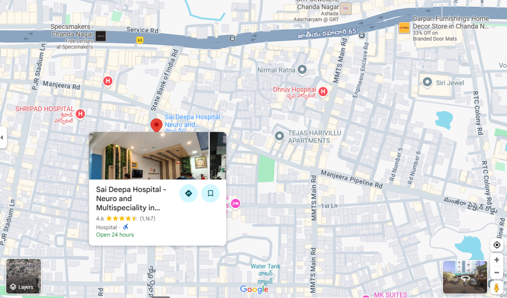
Sai Deepa Hospital - Neuro and Multispeciality
Plot no 387, Church road, Huda colony, Chanda Nagar, Hyderabad – 500050
Conclusion
The USG scan has transformed diagnostic medicine by providing a safe, quick, and cost-effective way to look inside the human body. From confirming pregnancies to diagnosing life-threatening conditions, its applications remain invaluable. Patients appreciate its non-invasive nature, while doctors rely on its accuracy and versatility.
As technology advances, ultrasonography will continue to play a crucial role in healthcare, ensuring timely diagnoses and better patient outcomes. Whether used in emergency medicine, obstetrics, or cardiology, the USG scan remains a window into the body’s hidden details – one that saves lives every day.


