What Is Digital X-Ray
- Home
- What Is Digital X-Ray
Digital X-Ray (State of the Art)
Medical imaging plays a crucial role in diagnosing and treating countless health conditions. Among the most common and reliable tools, the digital X-ray has become the cornerstone of diagnostic radiology. Unlike traditional film-based radiography, this advanced technology offers sharper images, reduced radiation exposure, and faster results, making it an indispensable part of modern healthcare.
Book Free Appointment
1L+
Happy Customers
25+
Qualified Doctors
50
Rooms
5000+
Successful Surgeries
Free
Consultation
24/7 Ambulance
Facility
Insurance
Claim Support
Evolution from Traditional X-Rays to Digital Imaging
Conventional X-rays relied on film processing, which often took time, demanded chemical use, and required physical storage. Patients had to wait for images to develop, and doctors often faced limitations in adjusting image quality. With the introduction of digital X-ray technology, radiology entered a new era.
This cutting-edge approach captures images electronically and processes them immediately on computer systems. Radiologists can enhance, enlarge, or manipulate images for more accurate analysis. Hospitals and diagnostic centers also benefit from digital archiving, which allows for quick retrieval of past reports without worrying about film deterioration.

Dr. Sasidhara Roa A
MBBS, MS
5000+ Successful Surgeries
11+ Years of experience
Dr. Sasidhara Rao A. is an experienced General and Laparoscopic Surgeon at Sree Sai Deepa Hospitals, Chandanagar, with over 11 years of expertise and 5000+ successful surgeries. He specializes in laparoscopic, laser, and microscopic surgeries, treating conditions like piles, fissures, varicose veins, and gallbladder issues.
Doctor’s Fellowships:
Fellowship - International Society of Coloproctology
Fellowship in Intimate Health
Fellowship in Diagnostic Endoscopy
Advantages of Digital X-Ray Technology
Digital radiography brings multiple benefits to both patients and healthcare providers.
1. Superior Image Quality
Digital X-rays deliver high-resolution images with excellent clarity. Physicians can zoom in, adjust contrast, and highlight specific areas, ensuring accurate diagnosis. Subtle fractures, infections, or tissue abnormalities often become more visible with these advanced imaging systems.
2. Reduced Radiation Exposure
One of the biggest advantages lies in the lower dose of radiation. Patients undergoing a digital X-ray are exposed to significantly less radiation compared to traditional methods. This makes it a safer option, especially for children, pregnant women (when medically necessary), and patients requiring multiple scans.
3. Quick Results and Better Workflow
Traditional X-rays required chemical processing, leading to delays. With digital radiography, images appear instantly on monitors, allowing doctors to provide faster diagnosis. This efficiency proves life-saving in emergency cases, such as trauma or suspected internal bleeding.
4. Eco-Friendly and Cost-Effective
By eliminating the need for films, chemicals, and darkroom processing, digital X-ray systems reduce environmental impact. They also save storage space, cut operational costs, and improve overall hospital efficiency.
5. Easy Storage and Sharing
Images captured digitally can be stored in hospital databases and shared electronically with specialists anywhere in the world. This seamless sharing encourages multidisciplinary consultation and enhances patient care.
Applications of Digital X-Ray in Healthcare
The versatility of digital radiography makes it an essential diagnostic tool across multiple medical fields.
1. Orthopedics
Doctors frequently use digital X-rays to examine fractures, bone deformities, and joint abnormalities. The precision of imaging helps surgeons plan orthopedic procedures and monitor recovery progress.
2. Dentistry
Dental professionals rely heavily on digital imaging for cavity detection, root canal assessments, and implant planning. Patients benefit from faster results and less radiation exposure.
3. Chest and Lung Examination
A digital chest X-ray remains one of the most common investigations for conditions such as pneumonia, tuberculosis, lung cancer, and COVID-related complications. The high-quality images allow radiologists to detect abnormalities with greater confidence.
4. Gastrointestinal Studies
Specialized digital X-rays with contrast studies help visualize the esophagus, stomach, and intestines. These scans assist in identifying ulcers, tumors, blockages, or swallowing disorders.
5. Cardiovascular Evaluation
In certain cases, digital radiography aids in assessing the size and shape of the heart, detecting fluid around it, and spotting vascular abnormalities.
6. Emergency Medicine
In trauma and accident cases, digital radiography delivers immediate imaging support to detect fractures, internal bleeding, or foreign bodies, enabling quick treatment decisions.
Get your surgery cost
Safety of Digital X-Ray Imaging
Many patients express concern about radiation exposure. However, digital X-ray technology significantly minimizes risks by using advanced sensors that require lower doses compared to film-based methods. Radiologists and technicians follow strict safety protocols, including protective shielding, to ensure patient safety.
For children and pregnant women, doctors carefully weigh the benefits against potential risks before recommending an X-ray. In most cases, the advantages of accurate diagnosis far outweigh minimal radiation exposure.
Digital X-Ray vs. Traditional X-Ray: A Quick Comparison
| Feature | Traditional X-Ray | Digital X-Ray (State of the Art) |
|---|---|---|
| Image Quality | Limited clarity, grainy films | High-resolution, adjustable images |
| Processing Time | Minutes to hours | Instant results |
| Radiation Dose | Higher exposure | Lower exposure |
| Storage | Physical films, prone to damage | Electronic, cloud-based storage |
| Cost Efficiency | Repeated films, higher costs | Long-term cost savings |
| Environment Impact | Requires chemicals and films | Eco-friendly, no hazardous waste |
This comparison highlights why digital X-rays represent the gold standard in modern diagnostics.
Accreditations

Saideepaneurocare Hospitals is NABH certified, a mark of excellence in patient safety and care. We follow stringent healthcare protocols and maintain world-class hygiene standards.

We are ISO 9001 certified, ensuring the highest standards in quality management and patient care. This certification reflects our commitment to efficient processes and continuous improvement in healthcare services.
Future of Digital Radiography
As technology continues to evolve, the future promises even more advancements. Artificial intelligence (AI) integration will help radiologists interpret images with greater speed and accuracy. Portable digital X-ray machines are already enhancing patient care in remote and rural areas. In the near future, imaging systems may combine with real-time telemedicine platforms, making expert consultations accessible globally.
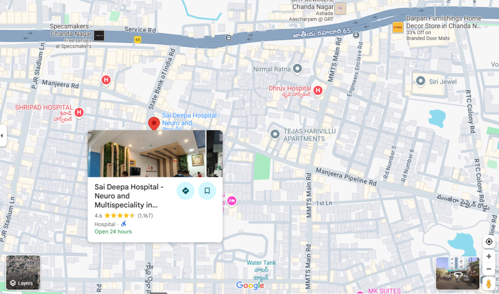
Sai Deepa Hospital - Neuro and Multispeciality
Plot no 387, Church road, Huda colony, Chanda Nagar, Hyderabad – 500050
Conclusion
The digital X-ray (state of the art) stands as a revolutionary advancement in medical imaging. With its superior clarity, faster processing, reduced radiation, and eco-friendly nature, it has redefined how healthcare professionals diagnose and treat patients. From routine check-ups to life-saving emergency care, digital radiography has become an indispensable tool in modern medicine.
Patients today can feel reassured knowing that their diagnostic journey involves safe, efficient, and precise imaging technology. As hospitals and diagnostic centers continue to embrace innovation, digital X-ray systems will remain at the heart of state-of-the-art healthcare delivery.


